Pool Season and Phlebitis: Recognizing Vein Inflammation Before Your First Swim in Ardsley
Published 2026-05-18

It's early June. The Ardsley Community Pool opens Memorial Day weekend, the Sprain Brook backyard pools are uncovered, and you've finally pulled the swimsuit out of the drawer for the first time since last August. You catch a glimpse of your calf in the mirror — and there it is. A reddened, ropy line along the inside of your leg, tender when you press it, warmer than the skin around it. Your first instinct is to cover it up. Your second instinct is to wonder if it's serious.
That ropy line has a clinical name. Vascular surgeons call it superficial thrombophlebitis, and the difference between a benign episode and something requiring urgent evaluation comes down to one thing: how deep the inflammation actually goes.
What Is Phlebitis?
Phlebitis is an inflammatory condition of a vein, most commonly affecting the superficial venous system in the legs. The inflamed vein develops a palpable, cord-like firmness, accompanied by redness, warmth, and localized tenderness along its course. When a blood clot forms within the inflamed segment, the condition becomes superficial thrombophlebitis, requiring duplex ultrasound to determine whether the clot has extended into the deep venous system.
Sound familiar? You're not overreacting — phlebitis warrants a clinical evaluation, particularly before summer activities that involve heat, sun exposure, and prolonged standing at the pool deck.
Why Westchester Residents Notice Phlebitis in Pool Season
There's a reason vascular surgery practices across Westchester see a spike in phlebitis presentations between Memorial Day and the Fourth of July. Three seasonal factors converge.
First, exposed skin. The vein was probably there in February, but a long-sleeved shirt and full-length pants kept it out of view. Pool season puts the leg under direct mirror scrutiny for the first time in months.
Second, dehydration. Warmer weather accelerates fluid loss. Dehydrated blood plasma thickens. Thickened plasma slows venous flow. Slowed flow within an already-dilated varicose vein creates the conditions for clot formation. The Ardsley resident who didn't drink enough water at the pool yesterday is at higher risk for inflammatory vein changes today.
Third, the heat itself. Vasodilation from sun exposure expands superficial veins, increasing pooling within already-incompetent vein segments. The combination of dilation, stasis, and minor trauma — a scrape against the pool ladder, pressure from a beach chair edge — is enough to trigger superficial thrombophlebitis in patients with underlying venous reflux.
Superficial vs. Deep: Why the Distinction Matters
The reason a vascular surgeon takes phlebitis seriously isn't usually the visible cord. It's what might be happening underneath.
Superficial thrombophlebitis affects veins close to the skin surface — typically the great saphenous vein or its tributaries. Most cases resolve with conservative management: graduated compression stockings, NSAIDs, warm compresses, and elevation. Symptoms improve within seven to fourteen days.
Deep vein thrombosis is a different clinical picture entirely. The clot sits within the femoral, popliteal, or calf deep veins, where dislodgement can produce pulmonary embolism. Approximately 10-20% of patients presenting with superficial thrombophlebitis are found, on duplex ultrasound, to have concurrent deep vein thrombosis. That ratio is exactly why "it's probably nothing" isn't an acceptable evaluation.
The vascular surgeon's job is to determine, definitively, which kind you have.
The Diagnostic Workup: What Happens at Our Ardsley Office
The evaluation for suspected phlebitis follows a structured sequence. The vascular surgeon doesn't guess. The vascular surgeon measures.
Step 1: Clinical Examination
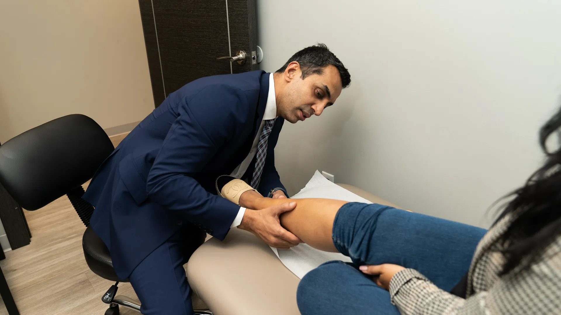
The physical exam maps the extent of the affected vein, palpates for cord firmness, assesses skin temperature, and checks for swelling above or below the inflamed segment. Swelling that extends into the calf or thigh raises the suspicion of deeper involvement.
Step 2: Duplex Ultrasound
Duplex ultrasound combines real-time imaging with Doppler flow analysis to visualize the affected vein in cross-section. The vascular surgeon can directly see whether a thrombus is present, how long it is, where it begins and ends, and — critically — whether it has propagated toward the saphenofemoral junction, the anatomic boundary between superficial and deep venous systems.
A thrombus within 3 centimeters of the saphenofemoral junction is treated as a deep clot. A thrombus several centimeters below it can typically be managed conservatively.
Step 3: Risk Stratification
Beyond imaging, the vascular surgeon considers personal history: previous DVT, recent immobilization, hormonal contraception, hypercoagulable disorders, recent surgery, and family clotting history. The combination of imaging findings and risk profile determines whether anticoagulation is necessary or whether compression and monitoring will suffice.
What Conservative Treatment Looks Like
For uncomplicated superficial thrombophlebitis, the vascular surgeon typically prescribes the following protocol:
- Graduated compression stockings, 20-30 mmHg, worn during waking hours for 3-4 weeks.
- NSAID therapy (typically ibuprofen 400-600 mg three times daily) for 7-10 days to reduce inflammation and pain.
- Warm compresses applied to the affected segment for 15-20 minutes, three times daily.
- Leg elevation above heart level for 20 minutes, three times daily.
- Walking — yes, walking. Immobilization worsens venous stasis. Movement activates the calf muscle pump.
Most patients see significant improvement within 7-10 days. The cord may remain palpable for several weeks as the inflamed segment remodels, but tenderness, redness, and warmth resolve much faster.
When Sclerotherapy or Underlying Treatment Comes Next
Phlebitis often signals that an underlying varicose vein or venous insufficiency exists. Once the acute inflammatory episode resolves, the vascular surgeon revisits the root cause. Untreated venous reflux makes future phlebitis episodes more likely. Definitive management — ultrasound-guided sclerotherapy, radiofrequency ablation, or VenaSeal closure of the refluxing saphenous vein — addresses the anatomy that produced the inflammation in the first place.
Read more: Can Varicose Veins Cause Blood Clots?
This is the difference between treating the episode and treating the disease. Schedule an evaluation at Vein Center Doctor in Ardsley.